There are 2 main sets of ligaments in the knee joint: the ‘Collateral Ligaments’, which run along either side of your knee joint, and the ‘Cruciate Ligaments’; Anterior Cruciate ligament (ACL) and Posterior Cruciate ligament (PCL) which sit inside your knee joint.
These ligaments hold the knee together and also provide the joint with stability and mobility to move.
Ligaments play a large role in bracing your knee joints for everyday activities such as walking, climbing, sitting or kneeling. When you injure or tear a ligament, you may feel as though your knees will not allow you to move or even hold you up.
The anterior cruciate ligament controls how far the Tibia (shin bone) can slide relative to the Femur. In other words, the ACL prevents too much forward movement.
The ACL can be torn from sudden pivoting movements to the knee joint. Football, soccer or netball are common sports, which have a high incidence of ACL injuries. Other activities include, losing control of your skis or falling off a ladder.
After an ACL is torn, if left untreated the knee can become quite unstable. You may experience episodes of your knee giving way, or buckling. The severity differs person to person. Instability can range from mild buckling with vigorous activity to severe buckling climbing stairs or attempting normal activities. It is the instability that leads a patient to surgical intervention.
The posterior cruciate ligament (PCL), centrally located behind the ACL, is less frequently injured.
When you injure your knee, all you know at first is that something is wrong. Injury to the ACL is like unraveling rope fibres. A partial tear can also occur but is rare. You can also injure other parts of the knee at the same time as you injure your ACL. Cartilages are at risk as is the gristle of the joint surface.
At the time of the injury you may feel a “pop” or “snap”. Onset of swelling and intense pain is usually immediate. You may also experience too much “play” in the joint.
Typically, the person is in intense pain and unable to continue with their activity. Immediate first aid is essential; ice, elevation and bracing the joint.
Seeking medical advice as soon as possible is advisable.
A medical examination by an Orthopaedic Surgeon helps us to determine the severity of your injury and your best treatment options. The earlier you are examined, the earlier your treatment and the better your chances for a successful recovery.
A knee ligament injury can be treated in 1 or 2 ways, non-surgically, or surgically. The choice depends on the severity of your injury and the level of activity to which you hope to return.
Rehabilitation will also be part of your treatment program.
There are a number of diagnostic tools in assessing an ACL tear.
Physical examination and history of the injury can specifically assess the amount of motion present and determine if the ACL is torn or not torn. It can also help to pinpoint the location of your problem.
Checking for abnormal motion in the knees and for swelling or tenderness is all part of this examination.
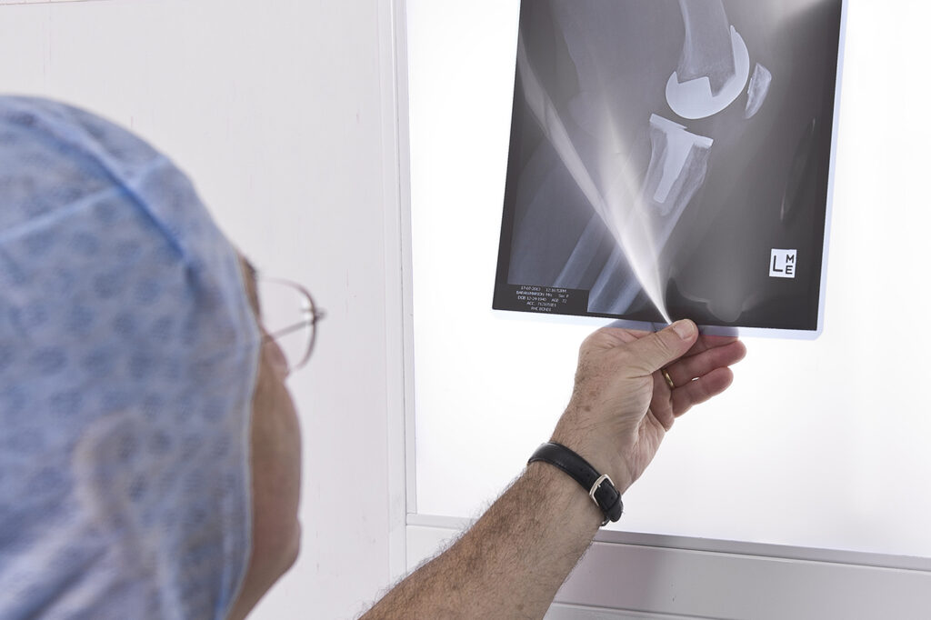
MRI, x-ray, Arthrogram and CT scans are used to verify a diagnosis if the physical examination is not conclusive.
The treatment options for each patient are individualized. Once Professor Kohan has made a diagnosis, together you can discuss which treatment option is best for you whether non-surgical or surgical, depend on many factors.
Treating your own injury without surgery is possible if no other tissue is injured and if your lifestyle will not put high demands on your joints. An athlete or more active person might need surgery to give the knee joint an extra edge against re-injury.
Rehabilitation follows either treatment option.
Non-Surgical Treatment
This treatment program includes a period of rest and exercise.
After injury, ice, elevation and support are used to control swelling. Using crutches or a brace helps you temporarily to rest your joint so that it can heal.
An exercise program to help return you to activity will be arranged. Strengthening your muscles to make up for your weakened ligament is a necessary long-term commitment.
Surgery
The most common type of surgery for an ACL injury is reconstruction. This involves replacing a torn ligament with a tendon (graft) from a hamstring. The graft is commonly fastened with screws.
To reconstruct your ACL, an arthroscopic technique is usually chosen. Surgery is followed by several months of rehabilitation to help restore your knee function.
After a diagnosis has usually been made, the patient will have to undergo an arthroscopy.
An arthroscopy provides a 100% accurate assessment of the type of injury that you have sustained in your knee then, based on this assessment an ACL or knee reconstruction will either take place at the same time or delayed to a later date.
An ACL reconstruction requires 2 puncture holes and a small incision. The small incisions are made to remove a portion tendon from your hamstring (called double loop semitendinosus and gracilis tendon graft). These grafts come from your hamstring and are is inserted into a dual hole and fixed in place with screws.
There will be a screw in your femur or thighbone and a screw in your tibia or shinbone. This technique provides the stability and future motion for your knee closest to that of an uninjured ACL.
Your incisions are closed with sutures, which need to be removed 10-14 days after surgery.
ACL reconstruction is considered a day only procedure. This means that you come into hospital a couple of hours before your operation and go home a couple of hours after the operation is completed.
When you wake up you may feel a bit groggy from the anaesthesia. You will have a bandage and knee brace on your knee.
Your recovery stay will be for approximately 45 minutes to 1 hour. Professor Kohan and his staff will monitor you, checking your blood pressure, temperature and pulse. Dr Kerr will also assess your pain level. Following this examination & if all ok you will then be able to go home.
Because the anaesthetic and pain medication may make you sleepy, arrange ahead of time to have someone drive you home.
A postoperative appointment is normally scheduled for a week to 10 days after the surgery.
Dressing
A compression bandage will be applied to your knee. Although it will be quite tight, the dressing itself is soft. This dressing is designed to absorb fluid, and/or blood. If the dressing becomes moist or blood stained, there is no need for alarm. You may change the dressing 2 days after the surgery, unless otherwise directed.
Once a dressing has been removed, cover the small cuts with bandaids.
Incisions
The incision is closed with sutures. These need to be removed 7-10 days after surgery. You may not get the incision wet until the staples are removed; therefore, you must sponge bath.
You may shower 2 days after the sutures are removed, but may not bathe or swim until 2 weeks from the surgery date. You may apply Vitamin E or moisturising lotion to the incision after the staples are removed. Some swelling and warmth is expected after surgery.
Medications
Pain management will begin in the Operating Room while you are still under anesthetic. At the time your knee will be extensively injected with long-acting local anesthetic and the drug Toradol. This should give you a very good pain control for about 20 hours.
Rest can help to relieve pain. In addition, prescription medications to take at home following your procedure may be required such as Panadine Forte or Nurofen.
Do not take Asprin for the first few days following surgery as it may increase bleeding.
Walking & Exercises
Upon discharge from the hospital you will be walking with crutches, and have a knee brace in situ.
Crutches may be needed for one to two weeks, or even longer.
Expectations
0 – 2 weeks after surgery your activity level is usually limited, however, you will be able to walk independently, use the bathroom, and perform normal activities of daily living.
2 – 4 weeks after surgery you will be able to engage in moderate activities, i.e. driving a car and climbing stairs. Your brace may be able to be removed in your home environment.
5 – 6 weeks after surgery you will have resumed most of your normal activities. The brace will be removed 100% of the time.
6 – 8 weeks after surgery complete surgical healing takes. During this time some swelling and discomfort is normal and should be manageable with prescribed medications. After this time the knee tissue begins to soften and become more natural.
Some patients may notice a small area of numbness on the lateral aspect (outside area) of the knee incision. This may or may not resolve over time.
A positive attitude and a commitment to your rehabilitation will give positive results.
Generally our ‘Estimate of Fees’ is accurate however, on occasion unforeseen circumstances can arise during the operation which may require additional medical services or a different, more costly prosthetic device to be used. If this happens there may be additional costs to you that are not covered by the estimate.
This will be fully explained to you after the operation should it occur.
Professor Kohan’s Surgical Fees
Medical Item No: 49542
Surgical Assistant Fees
The surgical assistant fees will either be billed to you directly or Professor Kohan will bill you on his behalf.
Medical Item Number: 51303
Anesthetic Fees
You will meet with Dr Kerr, the anaesthetist before your operation so that you can obtain an estimate of his fees. These will be billed to you directly.
Medical Item Numbers: 18225, 22045, 23111, 21402, 17620, 17690
There will be 2 ‘no charge’ consultations after your Total Hip Replacement procedure.
Aftercare appointments with Professor Kohan following your procedure include:
7 Days Post Surgery – This appointment is in order to check the skin cut & for Professor Kohan to asses your overall recovery.
14 Days Post Surgery – At this appointment the skin clips will be removed. An ultrasound will also be done by our radiographer in the rooms in order for Professor Kohan to check for blood clots.
6 Weeks Post Surgery – At this appointment Professor Kohan will asses the X-ray and monitor your overall recovery.
Consultations after this time attract a fee which is reimbursed in part from Medicare.
These fees should be discussed with the hospital directly. Please be sure to check with your health fund regarding a gap or out of pocket expenses.
If you are privately insured the prosthesis used in a Total Knee Replacement procedure is usually fully covered by your Health fund.
These fees are payable directly to the sonographer.

Dr. Farah’s specialty interests are hip and knee surgery, hip and knee arthroplasty (joint replacement), anterior minimally invasive hip replacement and sports knee surgery.