The word arthroscopy comes from two Greek words; Arthro – joint & Scope – to look. The term literally means to look within the joint.
Professor Kohan uses Knee Arthroscopy as a tool to diagnose mechanical problems within the knee and to surgically repair certain conditions, such as torn cartilage or ligaments.
Arthroscopy is a minimally invasive procedure with diagnostic and therapeutic merits.
Arthroscopic surgery is undertaken when conservative treatment options e.g. physiotherapy, anti-inflammatory medications & corticosteroid treatment have not provided adequate improvement in pain levels.
Arthroscopic surgery is an extremely valuable technique and is generally easier on the patient than open surgery. It is a minimally invasive procedure that is a reliable way to diagnose and correct knee problems.
Many patients have arthroscopic surgery as outpatients, either in a hospital or in a day surgery centre. They have the procedure early in the day, and leave in the afternoon or early evening.
The small surgical wounds from the arthroscopy, often not needing stitches, ensure a more pleasing appearance than the scars caused by open surgery. Because the wounds are small, the patient’s immediate post-operative pain is decreased. It is usual for patients to go back to work or school or resume daily activities within a few days.
Arthroscopy is useful in evaluating and treating the following conditions:
Meniscal Tears
These often are referred to as tears of the cartilage. The medial and lateral menisci are semilunar fibrous type shock absorbers, which are situated on either side of the knee. Tears in the meniscus are a common cause of knee pain. They sometimes cause intermittent swelling, clicking and catching.
Most tears have a degree of degeneration and occur in the part of the cartilage that does not have a blood supply. This means that they are not usually amenable to repair and require removal of the torn unstable fragments. As much of the meniscus is left in position as it does aid in the distribution of loads throughout the knee. A small number of cases, particularly those associated with a cruciate ligament tear occur at the periphery of the cartilage and may be able to be repaired.
If this is a possibility then the surgeon will discuss it with you prior to surgery.
Loose Bodies
Sometimes a piece of bone or cartilage can break off from the surface and cause intermittent locking of the knee. These can be easily removed at surgery.
Osteoarthritis
Some patients with this condition can benefit from a knee arthroscopy. Although this will not change the underlying wear and tear it can sometimes be helpful for acute mechanical symptoms, such as, catching, locking or intermittent swelling.
Chondromalacia of the Patella
This is a common cause of anterior knee pain and is often bilateral. Whilst most patients are treated conservatively some patients do benefit from using a motorised shaver to remove unstable areas of articular cartilage behind the knee cap.
Inflammatory Synovitis
Arthroscopy can be used to diagnose and sometimes treat this condition by arthroscopically removing the inflamed synovial lining.
Assessment of Cruciate Ligament Injuries
This diagnosis can usually be made prior to surgery. However, an arthroscopy can sometimes be helpful when diagnosis is not clear cut. An arthroscopy is usually performed in the initial stages of an anterior cruciate ligament reconstruction.
Local Cartilage Damage
Sometimes a piece of the articular cartilage can be sheared off from the end of the femur bone. This can cause ongoing pain due to the damaged surface. Many patients will benefit from arthroscopic debridement. Newer techniques such as chondrocyte grafting can be used.
This procedure can be used for growing a culture of cartilage that is taken from the patient and insertion of a gel like patch to cover the defect. This procedure is relatively new but the results reported so far are very promising. It does involve two operations to the knee, including a second open procedure.
An arthroscopic surgical procedure requires the use of a hospital operating room under general anaesthesia.
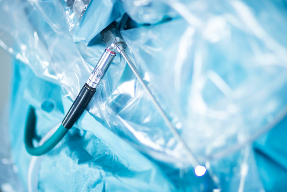
A small incision is made in the patient’s skin and a pencil shaped arthroscope is inserted with a miniature lens and light system that magnifies and illuminates the structures inside the joint.
This small instrument varies from 3mm to 5mm in diameter. Light is transmitted through fibreoptic cables to the end of the arthroscope that is inserted into the joints. By using a miniature television camera and screen combination, the interior of the joint is seen.
The television camera attached to the arthroscope displays the image of the joint on a television screen. The large image on the screen allows the joint to be seen directly to determine the extent of the injuries and then perform the particular surgical procedure if one is necessary.
Arthroscopic surgery is considered a day only procedure. This means that you come into hospital a couple of hours before your operation and go home a couple of hours after the operation is completed.
When you wake up you may feel a bit groggy from the anaesthesia. Professor Kohan and his staff will monitor you, checking your blood pressure, temperature and pulse. Dr Kerr will also assess your pain level. Following this examination & if all ok you will be able to go home.
Because the anaesthetic and pain medication may make you sleepy, arrange ahead of time to have someone drive you home.
A postoperative appointment is normally scheduled for a week to 10 days after the surgery.
A dressing will be applied to the incisions after your surgery. If the dressing becomes moist or blood stained, there is no need for alarm. You may change the dressing 2 days after the surgery, unless otherwise directed.
Once a dressing has been removed, cover the small cuts with bandaids.
The small surgical incisions are usually left open to allow drainage of fluid used during the surgery, but may on occasion be stitched. The point of entry may be sore and may develop bruising during the first few days after surgery. This bruising around the wounds will eventually disappear and does not require any special care.
Rest can help to relieve pain. In addition, prescription medications following your procedure may be required such as Panadine Forte or Nurofen.
Do not take Asprin for the first few days following surgery as it may increase bleeding.
Crutches may be required depending on your level of discomfort and the type of procedure undertaken. Crutches may be needed for one to two weeks, or even longer.
Physiotherapy treatment is also often required however this also depends on the type of procedure undertaken.
Generally our ‘Estimate of Fees’ is accurate however, on occasion unforeseen circumstances can arise during the operation which may require additional medical services or a different, more costly prosthetic device to be used. If this happens there may be additional costs to you that are not covered by the estimate.
This will be fully explained to you after the operation should it occur.
Professor Kohan’s Surgical Fees
Medical Item No: 49561 – 49557
Surgical Assistant Fees
The surgical assistant fees will either be billed to you directly or Professor Kohan will bill you on his behalf.
Medical Item Number: 51303
Anesthetic Fees
You will meet with Dr Kerr, the anaesthetist before your operation so that you can obtain an estimate of his fees. These will be billed to you directly.
Medical Item Numbers: 18225, 22045, 23111, 21402, 17620, 17690
There will be 1 ‘no charge’ consultation after your Knee Arthroscopy procedure.
Aftercare appointments with Professor Kohan following your procedure include:
5 – 7 Days Post Surgery – This appointment is in order to check the skin cut & for Professor Kohan to asses your overall recovery.
These fees should be discussed with the hospital directly. Please be sure to check with your health fund regarding a gap or out of pocket expenses.

Dr. Farah’s specialty interests are hip and knee surgery, hip and knee arthroplasty (joint replacement), anterior minimally invasive hip replacement and sports knee surgery.